Baby brain maps point to origins of neurological disorders

Roula Khalaf, Editor of the FT, selects her favourite stories in this weekly newsletter.
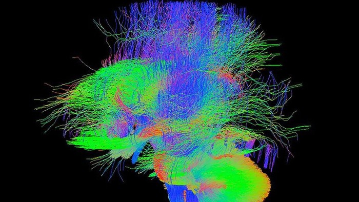
A remarkable brain-scanning project is under way in London. Over the next few years, researchers will generate high-resolution neural images of about 1,000 newborn babies and a further 500 foetuses in the womb.
This trove of 3D information about the wiring of the brain shortly before and after birth will help scientists understand the origins of neurological disorders such as autism and cerebral palsy — and should improve the care of babies who are born prematurely.
In this critical stage of early childhood development, the brain is growing very fast as trillions of connections form between its 80bn neurons. The way the brain develops at this time is influenced strongly by environmental factors, both positively — by intellectual and physical stimulus — and negatively — by disease, neglect or abuse.
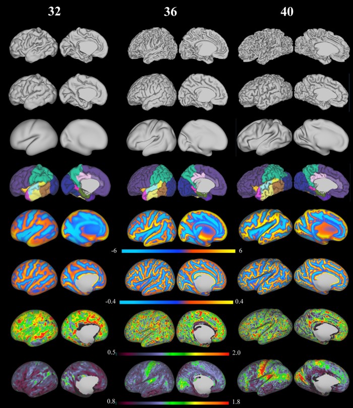
The Developing Human Connectome Project, led by King’s College London with €15m of funding from the European Research Council, uses magnetic resonance imaging (MRI) and computer analysis to take detailed pictures under circumstances that were previously impractical.
In adults, MRI scans require the subject to stay still for about 30 minutes, yet the King’s researchers, working with colleagues at Imperial College London and the University of Oxford, have adapted the technology to work on small, active babies even while they are still in the womb.
The project’s leader, Prof David Edwards, calls the connectome “a major advance in understanding human brain development”. It “will provide the first map of how the brain’s connections develop and how this goes wrong in disease”.

The first set of scans of 40 babies’ brainshas already been released for scientists around the world to download. Potential applications will expand greatly over the next few years as the MRI scanning programme hits its stride and the imaging information is linked to clinical, behavioural and genetic data about the infants whose brains have been scanned.
Elsewhere, other researchers are also using scanning technologies that were originally developed for adults to show what is going on inside babies’ brains. A team at the Massachusetts Institute of Technology’s McGovern Institute for Brain Research have designed an MRI machine with a reclining baby seat, a screen that allows the baby to watch videos, and space for a parent or researcher to sit inside the scanner. It also makes much less noise than typical machines.
The MIT neuroscientists used their machine to compare the brains of babies aged four to six months with adult brains. They showed the infants a series of films with different types of visual content, and found that baby and grown-up brains are organised in very similar ways. The same brain regions respond to faces and to outdoor scenes, for example, in babies and adults.
Although MRI provides much the most specific spatial resolution of neural activity, the cheaper and simpler electroencephalography (EEG) gives an indication of brain activity that is adequate for many studies. Using a soft skull cap lined with electrodes, which fits on the baby’s head, EEG records electrical signals in the brain.
Recent research using EEG has shown the importance of making eye contact with a baby — it helps to synchronise adult and babies’ brainwaves, and thus support communication and learning. Scientists at the University of Cambridge compared the neural activity of 36 infants with that of an adult singing nursery rhymes to them. Their brainwaves were significantly more synchronised when there was mutual eye contact. Infant “vocalisations” — attempts to communicate with the adult — increased, too.
“We don’t know what it is, yet, that causes this synchronous brain activity,” says Sam Wass, one of the Cambridge team. “Our findings suggested eye gaze and vocalisations may both somehow play a role, but the brain synchrony we were observing was at such high timescales [three to nine oscillations per second] that we still need to figure out how exactly eye gaze and vocalisations create it.”
A study at King’s College and University College London went further, combining MRI and EEG technology to investigate the bursts of high-amplitudebrainwaves that are vital for the healthy development of babies who are born very prematurely. This spontaneous firing helps to strengthen neuronal connections, forming the scaffolding that develops further with age.
Working with preterm babies 32 to 36 weeks after conception, the UCL researchers used EEG to identify electrical bursts, and MRI to pin down their source. It turned out that the bursts come from the insula, one of the most densely connected hubs in the developing cortex.
“This may offer new and exciting opportunities for monitoring how brain activity develops in preterm babies and a new understanding of how early irregularities can ultimately lead to disability,” says the study’s first author, Tomoki Arichi of King’s.
“Most research in early brain development focuses on structures instead of functions, so we’re hopeful that our methods can be used further to enhance our understanding of brain function before birth.”
Comments